7+ Gingival Anatomy
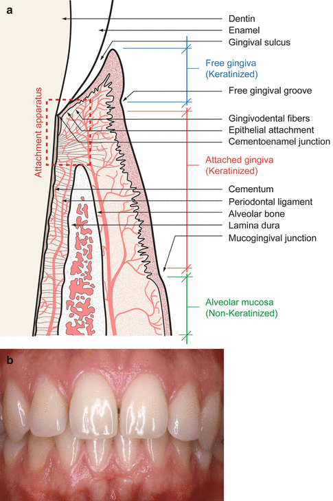
Laboratory Testing in Transfusion Medicine. The gingival sulcus is the area of space between the surface of the tooth and gingival tissue which surrounds it.
Fundamentals Springerlink
You need to be a group member to play the tournament.

. One descends to the skin of the chin Mental. Anatomy Histology of the Gingival Unit and Basic Oral Hygiene is a free dental continuing education course that covers a wide range of topics relevant to the oral healthcare professional. ISSN 2398-2942 Veterinary Clinics of North America.
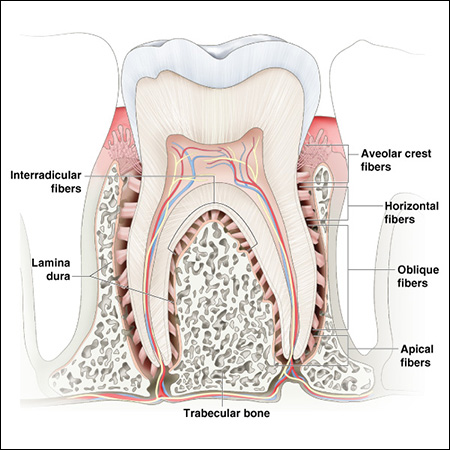
Small Animal Practice. 3 The alveolar mucosa is. The four components of the periodontium include the gingiva periodontal ligament cementum and the alveolar bone proper.
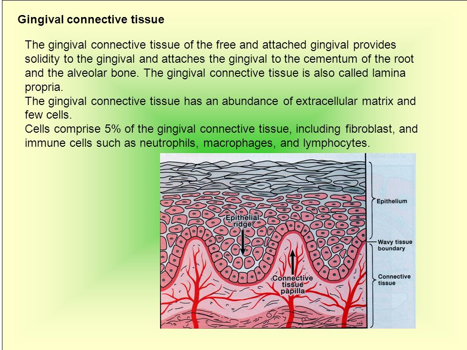
The gums also called gingiva include the mucosal tissue which cover the mandible and maxilla found inside the mouth. The sulcular epithelium lines the gingival sulcus. Mentalis emerges at the mental foramen and divides beneath the Triangularis muscle into three branches.
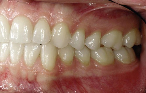
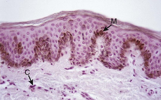
The tissues that surround envelop or embed the teeth including the gingiva cementum covering the tooth root periodontal ligament the supporting alveolar bone and the alveolar. Good gum health is important to both. Apical to the gingival sulcus is a band of highly permeable epithelium called the junctional epithelium which forms the epithelial attachment to the tooth.
Gingiva Gums Anatomy Head and Neck Gingiva Gums Contents Parts of Gum Structure Marginal Free Gingiva Gingival Groove Keratinized Gingiva Connected. Periodontal Anatomy Gingiva. This game is part of a tournament.
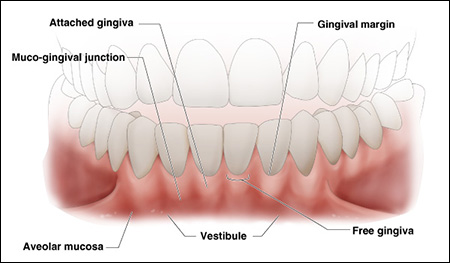
Located at roughly the same level as the bottom of the gingival sulcus. Attached gingiva is continuous with the free gingiva and is not movable as it is bound to the bone and cementum by connective tissue fibers. Attached gingiva tightly attached to underlying bone.
It lies between the free gingiva and the alveolar mucosa. Gum health and the. Periodontology Anatomy - Gingiva The gums or gingiva consist of the mucosal tissue which lies over the mandible and maxilla inside the mouth.
Join group and play Just play. The gingival margin or free gingival crest at the most superficial part of the marginal gingiva is also easily seen clinically and its location should be recorded on a patients chart. 7 The junctional epithelium is a.
The mental nerve n. Anatomy pathology deepening and elimination Odontol Tidskr. The gingival fibers are the connective tissue fibers which exist.
Anatomy Of The Periodontium An Overview Of Dental Anatomy Dentalcare
Periodontal Pocket
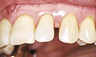
7 Reasons You Keep Getting Canker Sores Consumer Guide To Dentistry
![]()
Gingiva Types Histology And Clinical Aspects Kenhub
1 Anatomy Of The Periodontium Pocket Dentistry
Periodontology د باسم الاعسم Ppt Video Online Download

Periodontology Anatomy Gingiva
Anatomy Of The Periodontium An Overview Of Dental Anatomy Dentalcare
Periodontium With Its Components Gingiva Periodontal Fibers Pdl And Download Scientific Diagram
Smarty Pance Nccpa Eent Blueprint Review Course
1 Anatomy Of The Periodontium Pocket Dentistry
What Is The Healthiest Gum For Teeth Quora

The Anatomy Of The Gum Comparative Oral Ent Biology Openstax Cnx

Anatomy Of Gingiva And Um Pros Tho Don Tic Significance Pdf Dental Implant Human Tooth
Lecture Slides Anatomy Of Periodontium 2008
1 Anatomy Of The Periodontium Pocket Dentistry

Periodontium And Periodontal Disease Sciencedirect